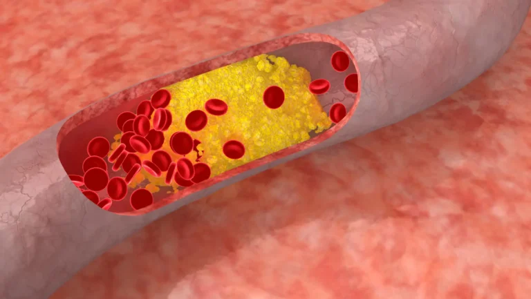
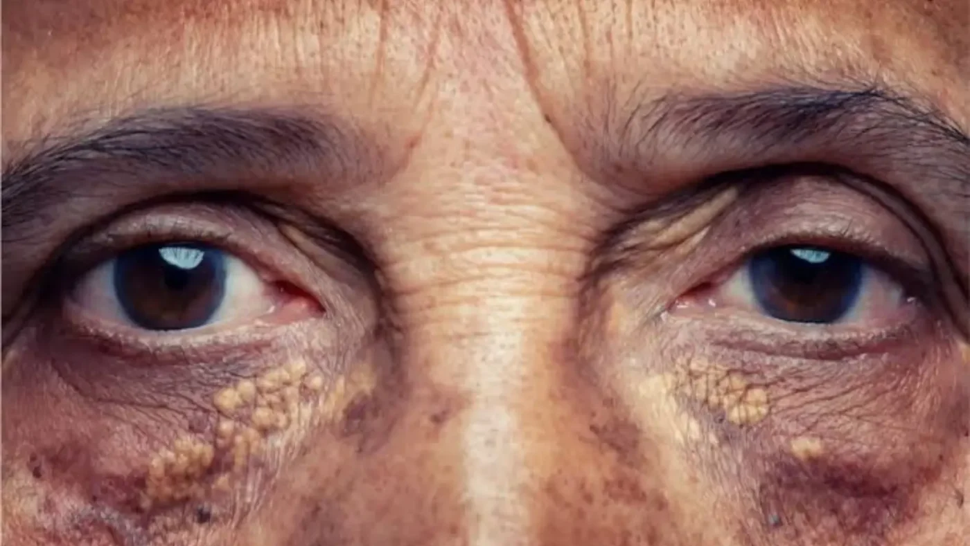

High cholesterol almost never causes symptoms, but a small group of people develop signs of high cholesterol on the face skin and around the eyes, mainly yellow eyelid plaques and a pale ring around the cornea. These markers, known medically as xanthelasma and corneal arcus, form when cholesterol-carrying particles leak into skin and eye tissue over years. The CDC confirms cholesterol itself stays silent, which is exactly why a visible clue on the face carries weight when it shows up.
This guide covers every confirmed facial marker, what each one looks like, when it points to a genetic condition, and what actually works to remove it.
Can High Cholesterol Really Show Up on the Face?
High cholesterol usually has no symptoms, and a blood test called a lipid profile is the only way to know your numbers. Facial signs appear in a minority of cases, and they cluster around specific situations rather than showing up randomly.
Signs of high cholesterol on face tissue show up more often in:
- People with cholesterol abnormalities lasting years, not a single high reading
- People with inherited disorders like familial hypercholesterolemia
- People with very high LDL for an extended stretch
- Older adults, simply from decades of lipid exposure
A facial sign alone proves nothing. About half of people with the most common marker, xanthelasma, have completely normal blood lipids. But it’s also not nothing. Doctors use it as a prompt to order bloodwork that might otherwise wait years.
Key takeaway: A facial sign is not proof of high cholesterol, but it can justify testing sooner, especially under age 50.
Xanthelasma: The Most Recognized Facial Sign of High Cholesterol
Xanthelasma is the most documented facial sign of high cholesterol, and dermatologists see it constantly in people between 35 and 55. It’s a flat, yellow plaque on the eyelid skin, and once you know what to look for, you won’t confuse it with anything else.
What Xanthelasma Looks Like
A soft, slightly raised, butter-yellow patch near the inner corner of the eye. It’s usually:
- On the upper eyelid first, sometimes spreading to the lower lid
- Symmetrical, showing up on both eyes around the same time
- Painless, with no itching or burning
- Slow-growing over months or years, not days
NIH’s StatPearls notes that xanthelasma affects 1.1% of women and 0.3% of men, typically between ages 35 and 55, most often on the inner upper eyelid.
Why Cholesterol Collects in This Area
Eyelid skin is thin and packed with tiny vessels near the surface. When LDL circulates at high levels for years, some particles leak out and get absorbed by immune cells called macrophages. Those cells swell with fat and become foam cells, the actual yellow material you see.
Can You Have Xanthelasma with Normal Cholesterol?
Yes, and this surprises most people. A ScienceDirect clinical reference states lipid levels are normal in roughly half of xanthelasma cases, though younger patients are more likely to have true hypercholesterolemia.
One study measuring lipid panels directly found that even with normal total cholesterol, 94% of xanthelasma patients had HDL well below the age-matched average, men averaging 31 mg/dL versus 45 mg/dL in healthy controls. A normal-looking panel can still hide a real HDL problem if nobody checks that number specifically.
Signs of High Cholesterol on Face
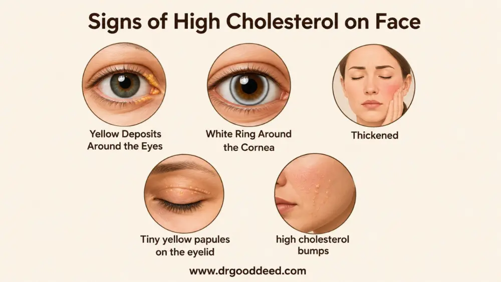
Xanthelasma isn’t the only clue. Several other signs of high cholesterol on face and eye tissue exist, each pointing to a slightly different mechanism.
Yellow Cholesterol Deposits Around the Eyes
Smaller, less defined yellowish deposits can appear near the lash line or under the eye. These are smaller versions of the same fat-and-macrophage process. They’re flat, don’t blanch under pressure, and don’t swell and fade like an allergic reaction would.
White Ring Around the Cornea
Corneal arcus is called arcus senilis in older age. It’s a thin, grayish-white ring at the edge of the iris, not the center, and it doesn’t affect vision. NIH data shows prevalence ranges from 20% to 35% depending on the population, and it’s present in nearly 100% of people over 80. In an elderly person, this white ring around the cornea mostly reflects aging vessels, not necessarily cholesterol risk.
Under age 50, the story changes. NIH notes corneal arcus in men under 40 is tied to greater cardiovascular risk, and the finding under 50, especially in men, warrants a lipid check because of the atherosclerosis link. A systematic review covering studies from 1960 through 2017 backs this: arcus in adults under 50 should prompt a lipid evaluation, though its absence doesn’t rule out heart disease.
Skin Changes Associated With Cholesterol Disorders
Severe, prolonged cholesterol elevation, almost always genetic rather than ordinary high cholesterol, can produce firmer, raised yellow-orange bumps called xanthomas elsewhere on the face or skin folds. These differ from eyelid xanthelasma by feeling firmer and appearing in clusters instead of one flat patch.
Less Common Facial Signs
A few rarer findings round out the list:
- Yellowish skin tone in primary biliary cholangitis, a liver disease that raises a specific lipid particle
- Tiny yellow papules on the eyelid margin in severe genetic lipid disorders
- Thickened, waxy eyelid skin in long-untreated cases
These are uncommon, but they show up in dermatology and lipid clinic case reports often enough to mention.
Treatment for Xanthelasma Around the Eyes
Once xanthelasma forms, diet and willpower won’t remove it. Treatment for xanthelasma around the eyes falls into four categories that differ widely in cost, recovery, and recurrence.
Cholesterol Management
Lowering LDL through statins or diet protects the heart and prevents new plaques, which is the real goal. It rarely shrinks existing ones. Controlling cholesterol matters for overall health, but existing xanthelasma rarely disappears from lipid management alone; physical removal is typically needed for cosmetic improvement.
Laser Removal
CO2 laser ablation has the strongest published results. A 2026 study of 295 patients tracked over 12 months found clearance above 99% for smaller lesions and around 95% for the largest, with recurrence at just 6.8%. A separate review of studies from 2021 to 2025 ranked CO2 laser as most effective overall, citing full resolution with a 13% to 16% recurrence rate.
Surgical Excision
Cutting the plaque out works well for large lesions, with one plastic surgery review reporting excellent outcomes when combined with eyelid surgery techniques. The tradeoff is longer recovery and real risk of scarring or eyelid pulling. Recurrence after excision ranges from 3% to 60%, depending on size and technique.
Chemical Treatments
Trichloroacetic acid, applied directly to the plaque, costs the least and needs no anesthesia, but results are unpredictable. One nearly three-year follow-up study found that 95% strength TCA led to recurrence or repeat treatment in 66.7% of patients, with success dropping from 70% at one year to 33% by the end of the study.
Risk of Recurrence
Across every method, new cholesterol keeps depositing in the same spot if the underlying lipid problem isn’t controlled. Pooled data puts overall recurrence between 25% and 60%, especially when hyperlipidemia stays uncontrolled. Removing the visible patch without fixing the lipid panel behind it treats a symptom, not the cause.
| Treatment | Clearance | Recurrence |
| CO2 laser | Up to 99%+ | 6.8% to 16% |
| Surgical excision | High for large lesions | 3% to 60% |
| TCA 70-95% | Variable, drops over time | 25% to 67% |
The Eye-Area Clues Often Confused With Cholesterol Signs
Not every bump near the eye is xanthelasma. These visible symptoms of high cholesterol get mixed up with three harmless skin conditions constantly.
Xanthelasma vs Milia
Milia are tiny, hard, white domes under the skin, usually grain-of-rice sized. They feel firm, unlike xanthelasma’s soft, flat texture. Milia form from trapped keratin and have no connection to the eyelid margin, while xanthelasma is a flat yellow plaque tied to cholesterol-rich cells.
Xanthelasma vs Syringomas
Syringomas show up as clusters of small bumps rather than one patch, and they’re flesh-colored, not yellow. They form from overgrown sweat ducts, usually below the lower lash line. NIH’s clinical reference lists xanthelasma directly among the standard look-alikes for eyelid syringoma.
Xanthelasma vs Normal Aging Changes
Eyelid sagging, fat bulges, and sun-thinned skin can all create shadows that look yellowish in certain light. None of these form the sharp-edged, flat plaque shape of true xanthelasma, and they won’t appear symmetrically the same way. A dermatologist can usually tell the difference by touch alone.
When Facial Cholesterol Signs Suggest a Genetic Cholesterol Disorder
Certain risk factors for visible cholesterol deposits combine into official diagnostic criteria for an inherited condition, not just a minor curiosity.
Familial Hypercholesterolemia and the Face
Familial hypercholesterolemia (FH) causes extremely high LDL from birth and affects roughly 1 in 250 people, though most remain undiagnosed. The Dutch Lipid Clinic Network criteria, the standard scoring system for FH, assigns 6 points for tendon xanthomas and 4 points for corneal arcus before age 45 toward a definitive diagnosis. A lipid specialty guideline confirms corneal arcus only counts toward FH if present under 45, and xanthelasma in a younger patient should also raise suspicion.
A 2025 case shows a 23-year-old woman with LDL at 509 mg/dL had corneal arcus, skin and tendon xanthomas, and joint deformities. Doctors confirmed homozygous FH using clinical scoring since genetic testing wasn’t available. Standard statin therapy failed to lower her LDL enough, so doctors added evolocumab, a newer injectable drug.
The result: an 82% LDL drop, from 509 to 89 mg/dL, with partial regression of her skin and tendon xanthomas. It’s a clear example of facial and skin signs leading to a serious diagnosis, and of aggressive treatment reversing both the numbers and the visible markers.
Warning Features That Deserve Prompt Testing
Get a lipid panel quickly if you notice:
- Corneal arcus before age 45, especially in men
- Xanthelasma before age 25
- Any eyelid plaque paired with a family history of early heart attack or stroke
- Tendon bumps, especially on the Achilles tendon or finger knuckles, alongside eye changes
Any one of these justifies bloodwork. Two or more together justify asking specifically about familial hypercholesterolemia screening.
What Facial Signs Cannot Tell You About Your Cholesterol
Facial signs can’t tell you your actual LDL, HDL, or triglyceride number. They can’t tell you how close you are to a heart attack. They can’t replace a lipid panel, the only direct measurement.
You can carry severely elevated LDL for years with a completely clear face. The 2026 update to US dyslipidemia guidelines now recommends checking lipoprotein(a), a separate genetic risk marker, precisely because so many high-risk people show no outward signs of high cholesterol on face or anywhere else. Absence of a facial clue means nothing about your real risk. Presence of one means it’s worth checking, not panicking.
FAQ
Can high cholesterol cause changes around the eyes?
Yes. Long-term elevated LDL causes xanthelasma, yellow eyelid plaques, and corneal arcus, a white ring around the cornea, both from cholesterol particles depositing in eyelid and corneal tissue over years.
What is xanthelasma and why does it occur?
Xanthelasma is a flat yellow plaque forming when LDL leaks through thin eyelid vessels and gets absorbed by macrophages, immune cells that swell with fat and create the visible deposit.
Does everyone with xanthelasma have high cholesterol?
No. About half of people with xanthelasma have normal cholesterol panels, though many still show low HDL specifically, so a full lipid panel is needed, not just a total cholesterol number.
What does a white ring around the cornea indicate?
After 50, it usually reflects normal aging vessels. In men under 40 to 45, it’s linked to higher cardiovascular risk and should prompt a lipid panel and broader risk check.
Is corneal arcus always related to cholesterol?
No. Past age 50, it’s present in most adults regardless of lipid levels. Before 45, especially in men, it’s far more likely tied to a genuine lipid disorder needing evaluation.
Can facial signs appear before cholesterol is diagnosed?
Yes. Xanthelasma and corneal arcus can appear years before a lipid panel, since high cholesterol produces no symptoms on its own and routine screening often gets delayed.
How is high cholesterol diagnosed?
Only through a blood lipid panel measuring total cholesterol, LDL, HDL, and triglycerides. Most healthy adults get tested every 4 to 6 years starting in early adulthood.
Can lowering cholesterol make xanthelasma disappear?
Rarely alone. Statins and diet changes prevent new plaques and protect the heart, but existing xanthelasma almost always needs laser, surgical, or chemical removal to actually clear.
Sources
- CDC: Testing for Cholesterol
- NCBI StatPearls: Xanthelasma Palpebrarum
- ScienceDirect: Xanthelasma Overview
- PubMed: Xanthelasma as a Clinical Indicator of Decreased HDL-C
- NCBI StatPearls: Arcus Senilis
- PMC: The Relation of Corneal Arcus With Cardiovascular Diseases, Systematic Review
- Lipid.org: Familial Hypercholesterolemia Screening and Diagnosis Executive Summary
- ClinicalTrials.gov: Dutch Lipid Clinic Network Criteria Protocol
- PMC: Corneal Arcus, Xanthomas, and Finger Deformities in Homozygous Familial Hypercholesterolemia
- Physicians Weekly: CO2 Laser Excision for Xanthelasma Palpebrarum
- JCAD: Treatment of Xanthelasma Palpebrarum Using Trichloroacetic Acid 80%
- NCBI: Syringoma StatPearls
- Family Heart Foundation: New US Dyslipidemia Guideline 2026
Disclaimer: This article is for general education only and does not replace a diagnosis from a doctor. Get a blood lipid test before assuming any skin or eye change is linked to cholesterol.