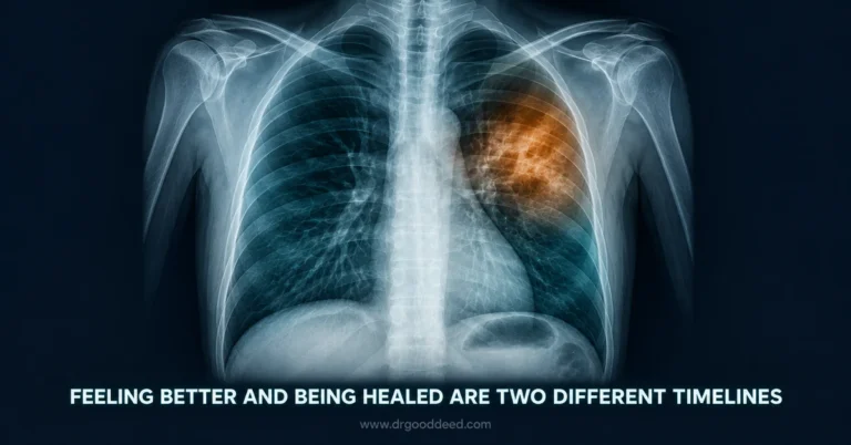
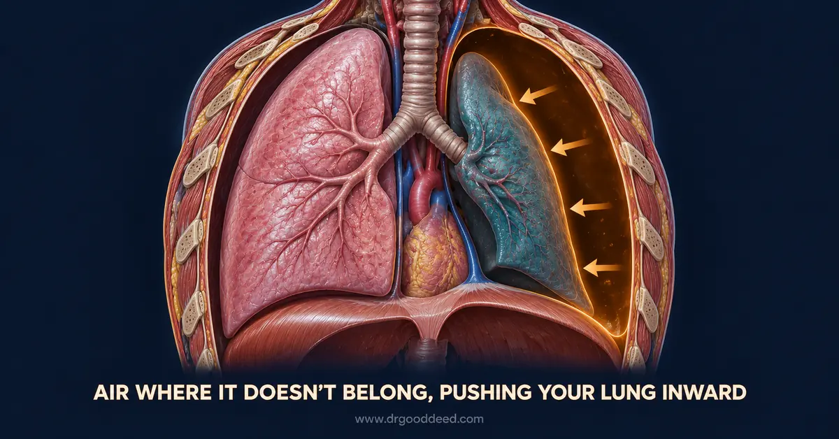

Sudden, sharp chest pain paired with breathlessness are the two hallmark collapsed lung symptoms doctors look for first. A collapsed lung, medically called pneumothorax, happens when air escapes into the pleural space and pushes on the lung tissue. Roughly 20 out of every 100,000 people in the US experience some form of it each year, and the presentation ranges from barely noticeable to life-threatening within minutes.
This guide covers collapsed lung symptoms, the real difference between pneumothorax and atelectasis, who faces higher risk, and exactly when sudden chest pain needs a 911 call instead of a wait-and-see approach.
What Is a Collapsed Lung?
A collapsed lung develops when air, blood, or fluid enters the space between the lung and the chest wall, called the pleural space, and compresses lung tissue from outside. The lung cannot fully expand once that pressure builds, and gas exchange drops.
Collapsed lung symptoms can appear within seconds after a chest injury or creep in gradually with an underlying lung disease.
- Pneumothorax is air specifically entering the pleural space. It can be total or partial, and it is the condition most people mean when they say “collapsed lung.”
- Traumatic pneumothorax follows a rib fracture, gunshot wound, stab wound, or a medical procedure like a central line placement or lung biopsy that accidentally punctures the pleura.
- Spontaneous pneumothorax occurs with no obvious external cause, usually from a ruptured air-filled sac (a bleb) on the lung surface.
- Iatrogenic pneumothorax, a subtype worth knowing, results from mechanical ventilation or needle-based procedures and is more common in hospitalized patients than most people realize.
Small pneumothoraces sometimes cause almost no symptoms at all, while larger ones can drop oxygen levels fast enough to become a medical emergency within minutes.
Common Symptoms of a Collapsed Lung
A sudden stab of chest pain and an inability to catch their breath.
- Sudden, one-sided chest pain that often feels sharp or stabbing rather than dull, typically worsening when you inhale deeply, cough, or move.
- Shortness of breath that ranges from mild breathlessness with a small pneumothorax to severe air hunger with a large one.
- Rapid, shallow breathing as the body compensates for reduced lung volume on the affected side.
- Rapid heart rate, since the heart works harder to maintain oxygen delivery when lung function drops.
- Bluish tint to the skin, lips, or nail beds (cyanosis), a late sign that signals dangerously low blood oxygen.
- Dry cough without mucus production, distinct from a cough tied to infection.
- Fatigue and lightheadedness, especially if the pneumothorax is large enough to affect blood pressure.
- Decreased or absent breath sounds on the affected side, a finding your provider checks with a stethoscope, not something you notice yourself.
Sudden Chest Pain
The chest pain of a collapsed lung usually starts abruptly rather than building gradually. It sits on one side, often described as sharp, stabbing, or like a knife between the ribs. Pneumothorax symptoms involving pain can radiate to the shoulder or back, which sometimes leads people to mistake it for a muscle strain or even a heart problem. Deep breaths, coughing, and certain positions typically make it worse.
Shortness of Breath
Breathlessness with a collapsed lung is not always proportional to how it looks from the outside. Some people with a small pneumothorax feel only mildly winded. Others with a large one struggle to speak in full sentences. Collapsed lung symptoms involving breathing difficulty tend to worsen with exertion and may not improve with rest, which is a useful distinguishing clue against anxiety-related breathlessness.
Other Possible Symptoms
A meaningful share of small pneumothoraces produce no obvious symptoms at all and get found incidentally on an X-ray taken for another reason. When symptoms do appear beyond the core two, they can include a fast pulse, clammy skin, anxiety tied to air hunger, and in severe cases, distended neck veins.
Expert Insight: A pneumothorax that feels minor at rest can decompensate quickly during exertion or air travel, because reduced cabin pressure lets trapped pleural air expand further. Clinicians generally advise against flying until follow-up imaging confirms the air pocket has resolved, even when the initial episode seemed mild.
Pneumothorax vs Atelectasis: What’s the Difference?
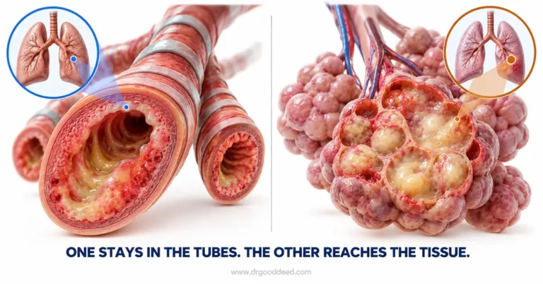
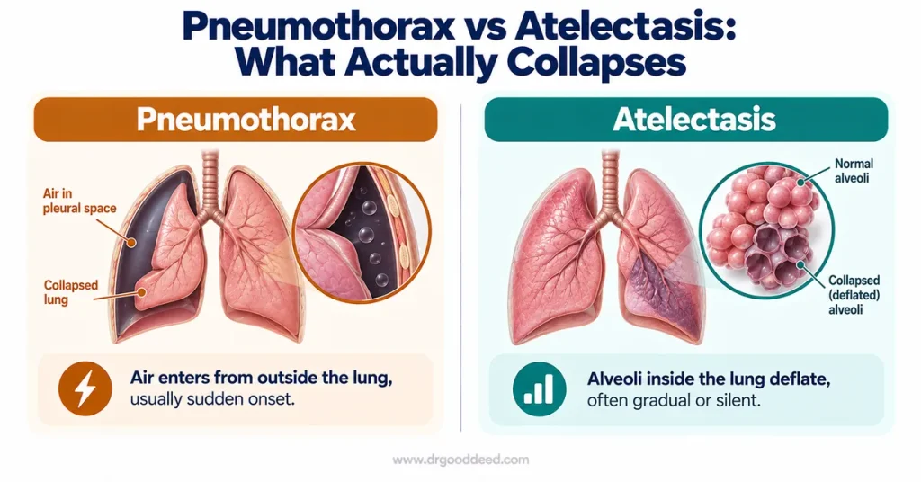
Pneumothorax means air has entered the pleural space from outside the lung tissue. Atelectasis means the air sacs (alveoli) inside the lung itself have deflated or collapsed, often without any air in the pleural space at all.
| Feature | Pneumothorax | Atelectasis |
| What collapses | The whole lung or a lobe, pushed inward by outside air | The alveoli themselves, deflating from blockage or pressure |
| Typical cause | Trauma, ruptured blebs, medical procedures | Surgery, mucus plugs, tumors, shallow breathing after anesthesia |
| Onset | Usually sudden | Often gradual, sometimes silent |
| Most common setting | Young, tall, thin men; chest trauma patients | Post-surgical patients, especially after abdominal or chest operations |
Spontaneous pneumothorax is one specific cause of atelectasis, not a synonym for it. Atelectasis is the single most common breathing complication after surgery, driven by shallow breathing, pain-limited coughing, and retained mucus rather than an air leak. Pneumothorax symptoms tend to hit abruptly and dramatically, while atelectasis frequently produces no symptoms at all in mild cases, only showing up on a routine post-operative chest X-ray.
What Causes a Collapsed Lung?
Air reaches the pleural space through one of three general routes: an external injury, a rupture from within the lung, or a medical procedure.
- Chest trauma, including rib fractures, gunshot wounds, stab wounds, and blunt force from car accidents or falls, tears the pleura and lets air in from outside.
- Ruptured blebs, small air-filled sacs on the lung surface, cause most cases of spontaneous pneumothorax in otherwise healthy young adults.
- Underlying lung disease, such as COPD, cystic fibrosis, or tuberculosis, weakens lung tissue and makes secondary pneumothorax more likely.
- Medical procedures, including central line placement, lung biopsy, and mechanical ventilation, can accidentally puncture the pleura.
- Sudden air pressure changes, from scuba diving or high-altitude travel, can rupture an existing bleb that had caused no prior symptoms.
- Catamenial pneumothorax, a rare cause tied to endometriosis, occurs when endometrial tissue in the chest cavity bleeds into the pleural space, typically around menstruation.
Who Is at Higher Risk?
Certain body types and habits raise the odds of a spontaneous collapse well above the general population baseline. Tall, thin individuals face a documented and often overlooked elevated risk, since the mechanical stress on lung tissue at the apex increases with height, making bleb rupture more likely even without any injury.
- Tall, thin body build, particularly in men between ages 20 and 40, due to greater mechanical stretch on the upper lung.
- Smoking, which raises spontaneous pneumothorax risk substantially compared with nonsmokers and also increases recurrence risk after a first episode.
- Existing lung disease, including COPD, asthma, and cystic fibrosis, which weakens lung tissue structurally.
- Family history, since primary spontaneous pneumothorax can run in families and, rarely, ties to a mutation in the FLCN gene.
- Recent chest trauma or thoracic surgery, which creates a direct entry point for air.
- Mechanical ventilation, which raises pressure inside already vulnerable lung tissue.
When Is a Collapsed Lung an Emergency?
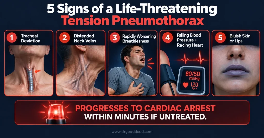
Tension pneumothorax happens when air keeps entering the pleural space through a one-way valve effect and cannot escape, building pressure that compresses not just the lung but the heart and major blood vessels. Left untreated, tension pneumothorax progresses to cardiovascular collapse and death, often within a short window.
- Tracheal deviation, where the windpipe visibly shifts away from the affected side, signals dangerously high pressure inside the chest.
- Distended neck veins, caused by blocked blood return to the heart, indicate the pressure is affecting circulation, not just breathing.
- Rapidly worsening breathlessness, especially if it escalates over minutes rather than staying stable, points toward tension physiology.
- Falling blood pressure with a racing heart, a combination that reflects the heart struggling to compensate for reduced venous return.
- Bluish skin, lips, or nails, a late but unmistakable sign of critically low oxygen that requires immediate action.
At the same time, small, stable pneumothoraces can and often do resolve with rest and monitoring alone, without any invasive procedure. Balanced awareness matters here: not every case of collapsed lung symptoms signals an emergency, but the specific combination above always does.
How Is a Collapsed Lung Treated?
Collapsed lung treatment depends almost entirely on size, symptom severity, and whether tension physiology is present. Emergency clinicians typically use a rough threshold of 2 centimeters of visible air on chest X-ray to separate small, observable cases from larger ones needing intervention.
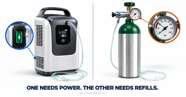
- Observation with oxygen, used for small, stable pneumothoraces under roughly 2 cm with minimal symptoms. Supplemental oxygen speeds pleural air reabsorption up to fourfold compared with room air, and the trapped air typically clears on its own within days to a couple of weeks.
- Needle aspiration, a bedside procedure using a large-bore needle to withdraw trapped air, reserved for larger or symptomatic cases in a stable patient.
- Chest tube (tube thoracostomy), inserted into the pleural space to continuously drain air, used for larger pneumothoraces, recurrent cases, or when needle aspiration fails.
- Needle decompression followed by chest tube, the immediate, non-negotiable response to suspected tension pneumothorax, performed before imaging confirmation because delay risks cardiac arrest.
- Pleurodesis or surgery (VATS), considered for recurrent pneumothorax, where the lung surface is sealed to the chest wall to prevent future episodes.
Recovery timelines vary widely. Someone with a small, observed pneumothorax may return to normal activity within one to two weeks, while a person recovering from chest tube placement or surgery, and adjusting to living without part of a lung in rarer severe cases, faces a longer recovery arc measured in weeks to months.
Pro Tip: Ask specifically whether your pneumothorax was measured using the Light index or Collins method on your chest X-ray report, since these size-estimation formulas directly determine whether your case qualifies for observation, needle aspiration, or a chest tube, and knowing the number helps you ask more informed follow-up questions.
When Should You Call 911 or See a Doctor?
| Situation | Action |
| Sudden one-sided chest pain plus shortness of breath, any severity | Seek prompt medical evaluation the same day |
| Chest pain or breathlessness after trauma, a fall, or a car accident | Call 911 immediately |
| Bluish lips or nails, rapid heart rate, or fainting | Call 911 immediately, this suggests tension physiology |
| Known lung disease with new, unexplained breathlessness | Contact your provider promptly; go to the ER if it worsens quickly |
| Mild breathlessness that has been stable for hours after a confirmed small pneumothorax | Follow your provider’s monitoring plan, but seek care if it changes |
Tension pneumothorax is rare relative to overall pneumothorax cases, but its speed of progression is exactly why sudden collapsed lung symptoms should never be shrugged off, especially in someone with chest trauma, a known lung condition, or a tall, thin build with no obvious cause.
FAQs
What does a collapsed lung feel like?
A sudden, sharp, one-sided chest pain that worsens with deep breaths, paired with breathlessness. Collapsed lung symptoms often feel like a stabbing sensation rather than pressure or tightness, distinguishing it from typical cardiac chest pain.
Can a collapsed lung heal on its own?
Yes, small pneumothoraces under about 2 cm often resolve without intervention. The trapped air reabsorbs at roughly 1.25% per day, with supplemental oxygen speeding this process up to fourfold.
Can you have a collapsed lung without knowing it?
Yes, small pneumothoraces and mild atelectasis frequently produce no noticeable symptoms and get discovered incidentally on a chest X-ray taken for an unrelated reason.
How long does it take to recover from a collapsed lung?
Small, observed cases typically resolve within one to two weeks. Chest tube or surgical collapsed lung treatment cases usually need several weeks to a few months for full recovery.
Is a collapsed lung life-threatening?
It can be. Most cases are not immediately dangerous, but tension pneumothorax specifically can cause cardiovascular collapse and death within minutes without emergency treatment.
Sources
- MedlinePlus: Collapsed Lung (Pneumothorax) link
- MedlinePlus: Collapsed Lung, Atelectasis, Pneumothorax link
- MedlinePlus Genetics: Primary Spontaneous Pneumothorax link
- Cleveland Clinic: Pneumothorax (Collapsed Lung) link
- American Lung Association: Pneumothorax Symptoms and Diagnosis link
- Northwestern Medicine: Atelectasis and Pneumothorax link
- Middlesex Health: Atelectasis link
- StatPearls (NCBI Bookshelf): Acute Pneumothorax Evaluation and Treatment link
- StatPearls (NCBI Bookshelf): Tension Pneumothorax link
- Emergency Medicine Cases: Management of Spontaneous Pneumothorax link
Medical Disclaimer: This article is for general information only and is not medical advice. It does not replace a diagnosis, treatment plan, or guidance from a qualified healthcare professional. Always talk to your doctor about your own symptoms and before starting, stopping, or changing any treatment. If you think you may be having a medical emergency, call 911 or go to your nearest emergency room.