
Medical Disclaimer: The blog is provided for informational and educational use only and should not be regarded as a substitute for professional medical advice, diagnosis, or treatment. You should consult a qualified neurosurgeon or your current doctor to obtain an evaluation and get specific recommendations concerning your condition. If you experience any worrying symptoms following surgery, seek immediate medical attention.
Neuroendoscopy is a minimally invasive surgical technique that helps neurosurgeons reach certain areas of the brain using a thin tube with a camera called an endoscope. It may be used to treat conditions such as hydrocephalus, selected brain cysts, and certain brain or pituitary tumors. Unlike some traditional brain surgeries, this approach can allow surgeons to work through small openings in the skull or through the nasal passages. The procedure, benefits, risks, and recovery time depend on the condition being treated and the patient’s overall health.
Understanding how neuroendoscopy works can help patients and caregivers prepare for discussions with their neurosurgeon about treatment options and expected outcomes.
Key Takeaways
- Neuroendoscopy is a minimally invasive surgical technique that uses a thin camera-equipped tube to reach selected areas of the brain through small openings.
- Neurosurgeons use this technique during different procedures, depending on the condition and the patient’s needs.
- It may help treat certain cases of hydrocephalus, brain cysts, and tumors.
- Benefits can include smaller incisions and less tissue disruption, but risks remain.
- Recovery varies with the procedure, the patient’s health, and any complications.
- Severe headache, repeated vomiting, seizures, or new confusion after surgery require urgent medical assessment.
| Condition | How the technique may help | Important note |
| Hydrocephalus | Endoscopic third ventriculostomy (ETV) creates an alternative pathway for cerebrospinal fluid. | Suitable only for selected patients. |
| Brain cysts | Allows access to certain cysts for drainage or treatment. | Depends on the cyst’s location and type. |
| Pituitary tumors | Provides access through the nasal passages in selected cases. | Requires specialist assessment. |
| Brain tumors | Helps surgeons reach or examine certain tumors near the ventricles or skull base. | Treatment depends on the tumor and its location. |
What Is Neuroendoscopy?
Neuroendoscopy is a surgical technique that uses a long, thin instrument called an endoscope to view and treat selected conditions affecting the brain and, in some procedures, other parts of the nervous system. A small camera and light at the tip help the surgeon see internal structures on a screen.
- This method does not need the large opening into the skull that is required by some conventional operations; instead, it makes use of small openings or natural pathways, such as the nose.
- It constitutes a kind of minimally invasive neurosurgery, but it is still surgery and therefore demands careful planning.
- Neuroendoscopy is not one specific operation. Neurosurgeons use it as part of different procedures, depending on the diagnosis, the location of the problem, and the patient’s overall health.
The surgeon considers imaging results, potential benefits, available alternatives, and individual risks before recommending this approach. Not every brain condition can be treated endoscopically.
Conditions It Can Treat
Neuroendoscopy may be used for selected cerebrospinal fluid disorders, brain cysts, and tumors. However, medical sources differ on some applications, so the appropriate procedure must be confirmed by a neurosurgeon.
Common uses include:
- Hydrocephalus: ETV may help selected patients by creating an opening that allows cerebrospinal fluid to flow.
- Brain cysts: Certain arachnoid or colloid cysts may be accessed and treated using endoscopic instruments.
- Ventricular tumors: The technique can help surgeons reach some tumors located within the brain’s fluid-filled spaces.
- Skull base tumors: An endoscopic approach may provide access to selected tumors near the base of the skull.
- Pituitary tumors: Surgeons may reach certain pituitary adenomas through the nasal passages.
The suitability of each approach depends on the condition’s size, location, and characteristics. Some patients need open surgery, medication, monitoring, or another treatment instead. The decision should be based on specialist assessment rather than the assumption that a minimally invasive procedure is always preferable.
How ETV Works
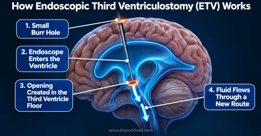
Endoscopic third ventriculostomy (ETV) is a procedure used to treat selected cases of hydrocephalus, a condition in which cerebrospinal fluid builds up inside the brain. It creates a new route for the fluid to flow, potentially avoiding the need for a shunt in some patients.
Steps of the Procedure
ETV is generally performed under general anesthesia. The exact technique depends on the patient’s anatomy and the surgeon’s assessment.
- Anesthesia: The patient receives general anesthesia and is monitored throughout surgery.
- Small opening: The surgeon makes a small opening, called a burr hole, in the skull.
- Endoscope insertion: A thin endoscope passes through the opening into a brain ventricle.
- Creating a pathway: The surgeon guides the instrument into the third ventricle and creates an opening in its floor.
- Widening the opening: A small balloon may be used to enlarge the opening when appropriate.
- Closure: The instruments are removed, and the surgical opening is closed.
The procedure may take about an hour, although the duration varies. The surgeon determines whether ETV is appropriate based on the cause of hydrocephalus and the patient’s individual anatomy.
EVT enables cerebrospinal fluid to pass through a new opening to regions where the body can absorb it, whereas a shunt works by diverting the fluid via a tube that has been implanted. Although shunts may become blocked or get infected, EVT too carries risks and might not be effective for all people.
Pituitary Tumor Surgery
In some cases, a surgeon is able to reach the pituitary gland via the nasal passages rather than by opening the skull; the endoscopic method enables access to the gland at the base of the brain.
- How appropriate this method is will depend on the size of the tumour, its position, and how it relates to adjacent structures.
- Surgery is not necessary in all cases of pituitary tumor, and no single approach can be used for every tumour.
- Patients must talk over the treatment plan with both their neurosurgeon and endocrinologist, as these specialists are able to outline the expected benefits, the possible effects on hormone levels, other treatment options, and the need for follow-up visits.
- Since recovery and the outcomes differ from person to person, it is more helpful to have individual advice than to depend on a general timetable.
Benefits Compared With Open Surgery
Neuroendoscopy can provide access to selected brain structures through smaller openings than some traditional operations. However, its advantages depend on the condition, the procedure, and the surgeon’s experience.
Potential benefits include:
- In some cases, the openings needed are smaller: certain procedures call for less extensive openings into the skull.
- There will be less disruption of tissue: this method could lessen the requirement to move or cut through the surrounding brain tissue.
- By using a camera, it is possible to see the surgical area with lighting.
- Access to the deeper structures: surgeons are able to reach certain areas which may be hard to access directly.
- Recovery might be faster: in some cases, patients will recover more quickly, according to the operation and their own health.
- The benefits cannot be guaranteed since neuroendoscopy has certain technical limitations and situations such as bleeding or poor visibility can make the procedure more difficult.
A systematic review carried out in 2024 by Minta and his colleagues looked at the use of ETVs and ventriculoperitoneal shunts in children suffering from hydrocephalus. The authors stated that both procedures can serve as reasonable first-line treatments since they have generally similar success rates, and added that more research is required in order to clarify the differences in complications. It therefore follows that ETV should not be automatically regarded as better than a shunt.
Risks and Success Rates
Even though neuroendoscopy is minimally invasive, it is still a form of brain surgery and may lead to serious complications. The extent of the risks will vary according to the procedure, the condition being treated, and the patient’s health.
Possible complications include:
- Experiencing bleeding either during or after the operation.
- Leakage of cerebrospinal fluid.
- Infection at the site of surgery or within the nervous system.
- Air gathering within the skull.
- There are fluid collections near the brain, such as a subdural hematoma or a hygroma.
- Injury to nearby blood vessels, nerves, or brain structures.
- Seizures or reactions to anesthesia.
For ETV specifically, a systematic review published in 2011 analyzed 2,985 procedures and reported an overall complication rate of 8.5%. It also identified uncommon but serious neurological complications. These historical figures cover ETV rather than every type of neuroendoscopy and should not be treated as an individual’s predicted risk.
Success rates also differ. Different definitions are used in the studies, for example those relating to improved symptoms, successful drainage of fluid, or avoiding the need for a shunt. It is not possible to reliably predict the outcome for one group of patients from the results obtained with another.
Ask your neurosurgeon about the expected benefit, the main risks in your case, the possibility of another procedure, and the alternatives if the operation does not work.
Recovery After Surgery
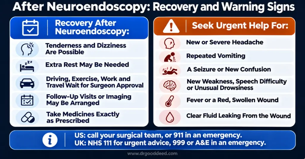
Recovery after neuroendoscopy depends on the operation performed, the patient’s general health, and whether complications occur. Your surgical team will determine when you can leave the hospital and return to normal activities.
During recovery, you may experience:
The area after surgery could be tender and your medical team might advise you to take pain relief.
- Dizziness: It is possible to feel lightheaded following surgery or anesthesia.
- You might need to get some more rest before going back to your normal routine.
- Activity restrictions: Driving, exercise, work, and travel may need to wait until your surgeon approves them.
- Follow-up visits: Your team may arrange examinations or imaging to check your progress.
The rate at which children and adults recover may vary. In some cases people feel better in just a few days, whereas in other cases it takes weeks or even longer. There is not a single recovery timetable that applies to all procedures.
Carefully follow your discharge instructions, take the medicines that have been prescribed exactly as instructed, keep the wound clean in accordance with the advice of your care team, and go to all your scheduled appointments. Do not stop or alter your medication without first consulting your clinician.
Before traveling by air or returning to strenuous activities, ask your surgeon when it is safe for you to do so.
When to Seek Urgent Help
Certain symptoms after brain surgery may indicate a complication and require prompt medical assessment. Contact your surgical team immediately if you notice concerning changes, even if the operation initially appeared successful.
Seek urgent medical advice for:
- A headache, either new or severe.
- Repeated vomiting.
- A fit or a new state of confusion.
- A new weakness, difficulty in speaking, or strange drowsiness.
- A fever or a wound that grows more red, swollen, or painful.
- Clear fluid leaking from the surgical wound.
If you have a shunt, redness or tenderness along the shunt, fever, headache, vomiting, or neck stiffness may indicate an infection or another complication. Contact your care team immediately. A suspected shunt blockage can also be an emergency.
If you are in the United States, get in touch with your surgical team for urgent advice or call 911 if it is a medical emergency. If you are in the United Kingdom, you should contact your surgical team or your GP, call NHS 111 for urgent advice, or else call 999 or go to A&E in the case of an emergency.
Frequently Asked Questions
Is neuroendoscopy major surgery?
That is a kind of neurosurgery, even if certain procedures involve smaller incisions than conventional operations, because the complexity and the recovery period depend on the condition being treated.
Is ETV better than a shunt?
Not always. ETV avoids an implanted drainage tube, but it is suitable only for selected patients and can fail. A neurosurgeon can explain which option is appropriate.
Will I have a visible scar?
Certain procedures leave behind a small scar on the scalp; however, when the surgeon accesses the pituitary gland via the nose there may be no obvious external scar on the scalp. The outcome varies according to the surgical approach.
Can children have neuroendoscopy?
Certainly. Endoscopic operations are employed by neurosurgeons in the treatment of children in certain situations, such as in some instances of hydrocephalus. Whether or not the procedure is carried out is based on the child’s age, the diagnosis, the anatomy, and the child’s general health.
Sources:
1.https://pubmed.ncbi.nlm.nih.gov/38010433/ link
2.https://pubmed.ncbi.nlm.nih.gov/22234198/ link
3.https://pubmed.ncbi.nlm.nih.gov/21631203/ link
4.https://pubmed.ncbi.nlm.nih.gov/40528870/ link
5.https://pubmed.ncbi.nlm.nih.gov/35103009/ link
Medical Disclaimer: This article is for general information only and is not medical advice. It does not replace a diagnosis, treatment plan, or guidance from a qualified healthcare professional. Always talk to your doctor about your own symptoms and before starting, stopping, or changing any treatment. If you think you may be having a medical emergency, call 911 or go to your nearest emergency room.